
Diagnostica e Radiologia Odontoiatrica
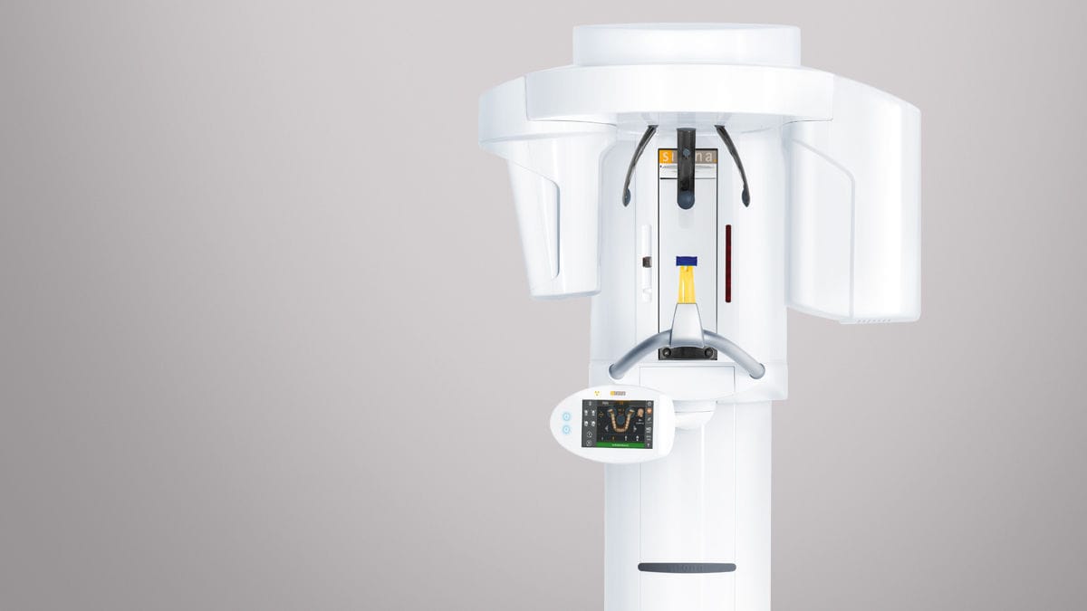
In the field of dental radiology, the Eminaj Dental Clinic is equipped with the technological tool of the moment: The Sirona 3D extraoral radiography system offers several versatile solutions for extraoral imaging needs. Combining the field of 3D technology with dedicated panoramic imaging, this affordable 2-in-1 offers the best of both worlds and offers the highest resolution and lowest radiation dose. The images allow doctors and dentists to quickly visualize teeth and bones of the skull, facilitating the diagnosis and clinical picture of the patient and the treatment plan to follow.
When and why to use dental radiography?
To clarify the patient’s clinical picture, the dentist takes x-rays that will be useful in formulating the diagnosis and the therapeutic plan
- Pre-implant Scenario
- Disorders of the dentition and malposed eighths
- Circumscribed or diffuse periodontal inflammation
- Cystic and similar lesions
- Osteo-fibro-cementitious dysplasia and thickening formations in general
- Paranasal sinuses, also in anticipation of surgery to lift the maxillary sinus floor
Executable imaging programs:
- Panoramic (panoramic, cephalometric, 3D)
- Segmented panoramic (panoramic, cephalometric, 3D)
- Maxillary sinus (panoramic, cephalometric, 3D)
- TMJ x 2 (panoramic, cephalometric, 3D)
- TMJ x 4 (panoramic, cephalometric, 3D)
- Lateral (cephalometric, 3D)
- Oblique (cephalometric, 3D)
- Frontal (AP / PA) (cephalometric, 3D)
- Submento-vertex (cephalometric, 3D)
- Carpus visualization (cephalometric, 3D)
- Digital archiving of images, recoverable even after some time
- Transmission of images via email

